Reconstruction of Calcaneal Fracture Malunion With Osteotomy and Subtalar Joint Salvage : Technique and Outcomes
2014-12-08 文章来源:上海同济大学附属同济医院 点击量:1211 我要说
Abstract
Background: The goal of this study was to discuss the outcomes of treating calcaneal fracture malunion by restoring the subtalar joint with a reconstructive osteotomy.
Methods: From May 2005 to November 2008, 24 patients (26 feet) with calcaneal malunions after a displaced intraarticular calcaneal fracture were treated by osteotomy and autogenous bone graft. The subtalar joint was preserved. The mean time from initial injury to reconstructive operation was 5.7 months (95% confidence interval, 4.5- 8.8 months). The displaced posterior facet was restored through a reconstructive osteotomy, whereas the bone defect in the calcaneus after reduction was filled with the exostosis that had been removed; iliac bone graft was used if necessary. All patients were evaluated clinically and radiographically at a minimum of 24 months. Twenty patients (21 feet) were followed for a mean of 34.2 months (29.0-39.4 months).
Results: According to American Orthopaedic Foot & Ankle Society (AOFAS) ankle and hindfoot score, the average score was 85.9 points (95% confidence interval, 81.5- 90.4 points), which was significantly higher than the preoperative assessment.Radiographs showed that Böhler’s angle,Gissane’s angle, talus declination angle, and width and height of calcaneus were improved to a great extent. Six patients had wound edge necrosis, and 2 had superficial infection. One patient required a subtalar fusion for subtalar arthritis at 2 years after surgery.
Conclusions: Restoring the subtalar joint with a reconstructive osteotomy and autogenous bone graft was an effective treatment method for selected calcaneal fracture malunions. It reconstructed calcaneal morphology and preserved the subtalar joint.
Level of Evidence: Level IV, retrospective case series.
Keywords: calcaneal fracture, malunion, subtalar joint, osteotomy, treatment, outcome
Nonoperative treatment of displaced calcaneal fractures, as well as inadequate reduction or failed surgery, may result in painful malunions with severe functional deficits that may seriously affect the patient’s quality of life. The typical deformities of calcaneus malunion include loss of calcaneal height, widening of the calcaneus, varus or valgus malalignment, and lateral shift with fracture dislocations.Residual incongruity of the posterior subtalar facet almost invariably leads to painful subtalar arthritis. The loss of calcaneal body height results in a more horizontal talus (loss of the talar declination angle),which may lead to anterior tibiotalar impingement and a decrease of ankle dorsiflexion,whereas malalignment may result in varus or valgus deformity of the calcaneus. Abnormal function of the ankle, subtalar, and transverse tarsal joints can lead to pain and disability.
Depending on the type of deformity, many authors have advocated different reconstructive strategies.Stephens and Sanders reported on a computed tomography(CT) classification system as well as a treatment protocol for calcaneal malunions. The classification system divided the calcaneal malunions into 3 categories: a type I malunion has lateral wall exostosis only, a type II malunion has lateral wall exostosis and subtalar arthritis, and a type III malunion has lateral wall exostosis, subtalar arthritis, and angular deformity. The core step of these protocols is reconstruction of the normal calcaneal morphology and subtalar arthrodesis. The symptoms of posttraumatic arthritis were alleviated through subtalar fusion. However, the talonavicular joint greatly influenced the overall hindfoot function.The subtalar joint also plays an important role in adduction and abduction of the hindfoot. After subtalar fusion, the foot maintained only about 50% of its transverse tarsal motion compared with the contralateral side.The subtalar joint also pronates and supinates during the gait cycle, which is essential for adapting to ground surface and structural abnormalities. A cadaver study by Savory et al showed that the motion of talonavicular joint decreased significantly after subtalar arthrodesis, including a 56% decrease in dorsiflexion and plantar flexion and a 70% decrease in inversion and eversion. Therefore, the ideal procedure to treat calcaneal malunion should not only correct the deformities but also preserve the motion and congruence of the subtalar joint.
We have found patients with malunited calcaneus fractures who still have normal articular cartilage in the subtalar joint. After the congruence of the subtalar joint is restored and the deformity is realigned, the symptoms caused by subtalar osteoarthritis can be greatly alleviated and a functional subtalar joint can be preserved. The purpose of this study was to report the short- to intermediate-term clinical and radiographic outcomes of this method.
Methods
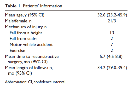
From May 2005 to November 2008, 62 calcaneal malunions in 51 patients were surgically treated in our department. According to preoperative evaluation and intraoperative direct visualization, 24 patients (26 calcaneus malunions) met the criteria and underwent reconstructive subtalar surgeries in this prospective study (Table 1).The rest of the patients excluded from the current study underwent reconstructive osteotomy with subtalar arthrodesis or other surgical procedures. Patients included in this study met the following criteria(1) the patient failed conservative treatment (2) the primary fracture was type I,II,or III according to Sanders malunion classification (type IV fractures were excluded) and (3) the patient had calcaneal malunions with mild to no osteoarthritis and the articular cartilage of the calcaneal posterior facet was near normal by radiographs, CT, and intraoperative visual examination.
There were 21 males and 3 females with an average age of 32.6 years (range, 20·52 years) in this group. Among them, there were 2 bilateral malunions and no open fracture malunions. All patients had been initially managed at other hospitals, including 22 treated conservatively and 2 surgically (closed reduction and Steinmann pin fixation). Comorbidities included cardiac disease in 1 patient, hypertension in 4, and smoking in 2.The average period from injury to reconstructive operation was 5.7 months (95% confidence interval [CI], 4.5·8.8 months). All patients had pain, walking disability, loss of the longitudinal arch, and anterior tibiotalar impingement as their main complaints. Twenty three of the 26 feet (88.5%) had severe symptoms that seriously affected daily life or activity. Twenty four patients (26 feet) underwent the reconstructive surgeries,including 19 feet with iliac crest autograft and 7 feet with the excised lateral exostosis. In this series,18 feet had a tongue osteotomy.Twenty four follow-up assessments were carried out at 1 year and 2 years post operation.Twenty patients (21 feet) were followed for a mean of 34.2 months (range, 29.0-39.4 months).
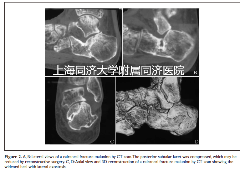
Preoperatively, radiographs including lateral and axial views of the calcaneus and mortise view of ankle joint (Figure 1) as well as CT scans and 3·D reconstructions were taken to evaluate the deformities (Figure 2).
Surgical Technique
Under epidural anesthesia, the operation was performed with the patient in a lateral position. A thigh tourniquet was inflated.We used a standard lateral extensile L-shaped approach to expose the calcaneal wall;3 Kirschner wires were placed into the lateral malleolus,cuboid, and talus neck.
An osteotomy was performed using a combination of a saw and an osteotome.The osteotomy started from posterior to anterior along the longitudinal axis of the calcaneus. The width deformity of calcaneus was corrected, and the area of impingement in the subfibular region was decompressed.Usually the thickness of the excised lateral wall was estimated and measured by preoperative axial radiographs and intraoperative visualization.The exostectomy sometimes continued to the level of the calcaneocuboid joint so as to decompress the calcaneocuboid joint.

After removal of the lateral exostosis, the depressed posterior facet could be seen. The excised lateral wall fragment was maintained in 1 piece, and the surfaces were shaved to proper shape for later use as a bone block autograft. According to preoperative CT scans and direct intraoperative visualization, a decision was made whether the subtalar joint should undergo arthrodesis or reconstruction. If the cartilage surface was almost normal, or at least two thirds of the cartilage surface was normal, especially the central cartilage surface, a reconstructive procedure was chosen.
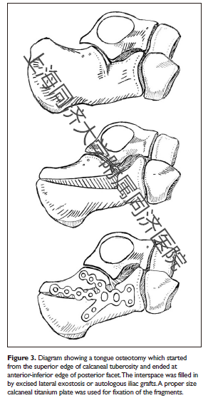
The typical primary fracture line usually lies in the sagittal plane and runs through the posterior facet of the subtalar joint.The collapsed and rotated posterior articular facet fragment that had been malunited was separated again along the primary fracture line with an osteotomy.With distraction, the body of the calcaneus was pulled plantar ward with respect to the medial aspect of the talus to reestablish the height.With a C arm fluoroscopy,a lateral view was acquired to verify the height that needed to be corrected,while an axial view was used to verify the angle needed to reduce varus/valgus deformity.According to our experience, the most posterior superior part of the tuberosity usually did not need to be completely removed but was kept connected to the calcaneal thalamus.The depressed posterior articular facet fragment was elevated upward and backward to restore the articular congruence.If the malunion had a varus or valgus deformity,the tuberosity was distracted in the opposite position to restore the alignment.If the primary fracture line was difficult to identify,we used a tongue osteotomy (Figure 3).After the depressed facet was identified,the osteotomy started from the superior edge of calcaneal tuberosity and ended at the anterior inferior edge of the posterior facet.Thus,the calcaneal posterior facet with the superior border of the calcaneus formed a tongue fragment.We distracted the tongue fragment upward and backward to restore the congruence of the subtalar joint in this subset of patients.Reducing and elevating the posterior facet usually left a defect in the cancellous bone beneath the facet.The fragment excised from the lateral wall was used to graft and support the facet fragment while holding the hindfoot in distraction and slight valgus. If a large defect appeared after the posterior facet elevation, further autogenous tricortical bone graft was taken from the iliac crest.Thus,the subtalar joint was reconstructed as well as the anterior process,Böhler’s and Gissane’s angles,and the calcaneocuboid joint.
After rebuilding the calcaneal morphology,a proper size calcaneal titanium plate was placed on the lateral wall.The elevated posterior facet fragment should be fixed by 2 or 3 cortical screws.Several cortical lag screws (3.5 mm) were inserted into the sustentacular fragment to maintain the reduction of the posterior facet.In most cases, all screws were placed through the plate holes to keep the fragments stable. Screws can also be placed outside the plate if necessary.C arm fluoroscopy was used to determine the proper reduction and screw placement.Care was taken to avoid varus or notable valgus of the calcaneus on the axial view.
The wound was closed in 2 layers with interrupted subcutaneous and skin sutures over a suction drain,which was placed so that it exited at the proximal tip of the vertical limb of the incision.In some cases when the thalamus was elevated over 1.5 cm,it was difficult to close the incision. Instead,the surgeon performed a skin flap transfer and dermatoplasty (2 of the 26 feet).
Postoperative Management
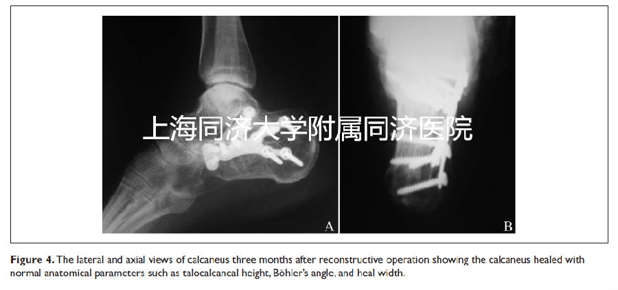
The patients were not immobilized in a plaster cast and were encouraged to move their toes and ankle 2 days postoperatively. Weight bearing was delayed for 10 to 12 weeks,until fracture healing was confirmed by radiological evaluation (Figure 4). Patients were reviewed at 6 weeks,3 months,6 months,1 year, and 2 years after operation,with clinical and radiographic assessment of the progress of healing and complications. The motion of the subtalar joint was assessed by physical examination.The operated foot was evaluated with the American Orthopaedic Foot & Ankle Society (AOFAS) ankle and hindfoot score before the operation and at 1 year and 2 years after the operation. Patients were also evaluated by weight bearing anteroposterior, lateral, and mortise radiographs of the ankle; weight bearing anteroposterior and lateral radiographs of the foot; and axial radiographs of the calcaneus.
Statistical Methods
Data are shown as the median and range, the mean and 95% CI, or the mean and standard deviation (SD) and are compared with paired t tests. A P value of less than .05 was considered to be significant with use of a 2-tailed test. All statistical analyses were carried out with SAS 9.1.3 (SAS Institute Inc, Cary, NC).
Results
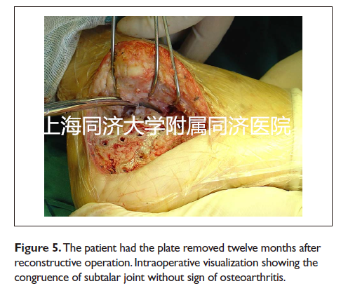
The average time to union was 11.2 weeks (95% CI, 10.8·12.7 weeks). Partial weight bearing was started at 8 weeks postoperatively, whereas full weight bearing was started at an average of 12.2 weeks (95% CI, 12.5·14.0 weeks). At 1 year of follow up, 5 (23.8%) patients had moderate pain and 8(38.1%) patients had mild pain occasionally; most of the pain was on the lateral aspect of the ankle (11/13).Six (28.6%) patients still had partial loss of subtalar joint motion; 5 of them believed that it did not interfere with their daily activities. At 2 years of follow up, only 3(14.3%) patients still had moderate pain, and 6 (28.6%) patients had mild pain occasionally. Three (14.3%) patients still had partial restriction of motion, but none of them believed that it interfered with their daily activities. Seventeen (85%) patients were satisfied with the results and returned to their preinjury work and activities, 2 (10%) patients had changed their occupation and worked part time, and 1 did not resume work. Eighteen (85.7%) patients had a stable hindfoot subjectively and clinically. Two (9.5%) patients had a slight limp when walking. One patient had ongoing hindfoot pain and restricted movement and underwent subtalar arthrodesis at 2 years after our reconstructive operation. Eighteen (85.7%) patients had no discomfort or stiffness when passively exercising the subtalar joint, whereas 2 patients (9.5%) felt stiff and 1 patient (4.8%) felt stiff with pain. The average AOFAS score was 85.9 points (95% CI, 81.5·90.4 points), whereas the average score preoperatively was 29.8 points (95% CI, 23.2·36.4 points) (Table 2). An almost normal subtalar articular surface was seen intraoperatively when removing the plate (Figures 4 and 5).
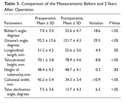
According to radiographs taken 2 years postoperatively, the calcaneal morphology was much closer to anatomic status. The average Böhler’s angle was 25.6 ± 4.7 degrees, the average Gissane’s angle was 121.7 ± 4.3 degrees,the average calcaneal length was 55.6 ± 3.6 mm,the average talocalcaneal height was 78.9 ± 4.6 mm,the average height of tuberosity was 48.7 ± 4.1 mm,the average calcaneal width was 34.3 ± 3.4 mm,and the average talus declination angle was 13.7 ± 4.3 degrees.The comparison of differences before and after operation in radiographic images is summarized in Table 3. There were significant differences between the 2 groups in Böhler’s angle, Gissane’s angle,talocalcaneal height,calcaneal width,and talus declination angle.The morphologies of most feet were well restored and the patients could wear normal shoes eventually. Of the 6 patients with radiographic arthritis, 1 had mild pain and 1 had continuous pain.The remaining 4 were asymptomatic.Six patients had wound edge necrosis and 2 had a superficial infection.One patient required a subtalar fusion for subtalar arthritis at 2 years after surgery.

Discussion
Nonoperative treatment of calcaneal fractures may result in malunion,which may result in long lasting disability for most patients.Many authors have described salvage measures for this problem.Braly et al reported using lateral wall osteotomy without subtalar arthrodesis as a treatment alternative for malunited calcaneal fractures. Nine of their 11 patients had satisfactory results at an average follow up of 28 months. Carr et al described a technique of restoring the height of the hindfoot by distraction of the subtalar joint with bone block arthrodesis. Several complications were associated with this procedure, including nonunion, superficial wound problem, broken screws, and varus position.The current mainstream techniques for calcaneal malunions include lateral wall decompression, in situ subtalar arthrodesis, distraction bone block arthrodesis, corrective osteotomy and subtalar arthrodesis, and triple arthrodesis. Distraction bone block arthrodesis is indicated in patients such as Stephens type II malunions and Zwipp type 3 malunions, whereas for more severe deformity (Stephens type III, Zwipp types 4 and 5), the subtalar joint can undergo arthrodesis with corrective calcaneal osteotomy.The goal of these measures is correction of the deformity and relief of the pain by osteotomy and subtalar arthrodesis.However,there were few reports about restoring subtalar congruency with reconstructive osteotomy when treating calcaneal malunions.
The Stephens and Sanders classification is simple and easy to use,but it does not cover all types of deformities. Traumatic osteoarthritis in the subtalar joint correlates not only with the severity of the injury but also with the duration of time between primary injury and surgery.There are inconsistencies between patients’complaints and severity of traumatic osteoarthritis. For some types of fractures, the fracture line of the malunions after removal of the lateral wall exostosis can be clearly visualized if the time since fracture is less than 12 months and almost normal articular cartilage surface is found.The longest period of the cases in this study was 24 months.This patient presented an almost normal cartilage surface (Sanders type I fracture,Stephens Sanders type II malunion) on radiographs, and no sign of subtalar osteoarthritis was visualized during the operation.
Treatment of any hindfoot deformity should include correction of the deformity and preservation of complex hindfoot motion. The movements of the subtalar complex are fundamental to transferring the forces from the foot to the leg during the early part of the stance phase of gait. Mann and Baumgarten reported a 50% loss of forefoot abduction and adduction after an isolated subtalar arthrodesis. Savory et al found that the range of motion of the talonavicular joint changes significantly after isolated subtalar joint arthrodesis. In their research, there was a 56% loss of dorsiflexion and plantar flexion at the sagittal plane and a 70% loss of eversion and inversion in the coronal plane. The investigators were convinced that normal range of subtalar joint motion is essential to the talonavicular joint.
Indications and Techniques of the Reconstructive Surgery
It’s appropriate for patients with minimal arthritic change at the subtalar joint on radiographs to have reconstructive surgery. Patients should be less than 12 months out since the fracture. A malunion caused by a Sanders type I to III fracture is suitable for this osteotomy. A Sanders type IV fracture is not suitable because of the severely damaged cartilage articular facet. The calcaneus of the patients should have a compressed posterior facet that can be clearly seen on preoperative radiographs or CT scan. The severity of osteoarthritis and the articular facet was evaluated by radiographs and CT; magnetic resonance imaging could also provide useful information. Intraoperative visualization is of great importance.
The most obvious benefit of this surgery is the salvage of subtalar joint instead of arthrodesis. Thus,the motion of subtalar joint is preserved to a great extent,which may reduce the stress on the adjacent hindfoot joints and reduce the later development of degenerative arthrosis in these joints.Our study included 18 feet (85.7%) that had no discomfort or stiffness when patients passively exercised the subtalar joint; only 2 (9.5%) felt rigid and 1 (4.8%) felt rigid with pain.
Gavlik et al considered corrections of the deformed Böhler’s angle, Gissane’s angle, heel width, and weight bearing axis of calcaneus as the criteria of an accurate reduction.If a calcaneal fracture malunion has mild osteoarthritis, unacceptable deformity still needs to be corrected by surgery.
Several incisions have been described for calcaneal fracture malunions, varying according to different surgical procedures. In this group of patients, to expose the subtalar joint and the calcaneal lateral wall, a lateral extensile L shaped approach was used. The depressed part of posterior facet could be fully visualized through this incision. The wound closure was not too difficult, as the lateral wall osteotomy relaxed the flap somewhat and the flap had reasonably good elasticity. In some cases when the thalamus was elevated more than 1.5 cm and a larger bone graft was used, it was difficult to close the incision. The surgeon performed a skin flap transfer and dermatoplasty in 2 feet of the 21 feet in our group, and all the flaps survived. This procedure requires microsurgery techniques.
The reduced posterior facet should be fixed and prevented from collapsing post operation by using a plate. There were no implant failures in this series. This plate (designed by G.R. Y.) is widely used to treat fresh displaced calcaneal fractures in China. We believe that the best procedure to prevent implant failure is to correct the malunion with appropriate autogenous bone grafts, leaving no defects.
This technique has the risk of subtalar osteoarthritis in the long term, which may require an arthrodesis eventually. Melcher et al followed 16 patients with intra articular calcaneal fractures for more than 10 years. In no case was there indication of need for a secondary arthrodesis. Although most patients showed slowly progressing posttraumatic subtalar osteoarthritis on radiographs, the subjective results (pain, capacity to work, sports participation) at 10 years were clearly better than at 3 years after surgery. In our group, radiographs showed degeneration in 6 patients, but only 1 patient had pain and discomfort. Once the deformity of the calcaneus is corrected by reconstructive surgery, a supplementary subtalar arthrodesis would be much easier if necessary.
Conclusions
There are different procedures for treating calcaneal fracture malunions, and the results of our study found that restoring the subtalar joint with reconstructive osteotomy and autogenous bone graft gave satisfactory outcomes and a low reoperation rate for selected patients. Through our surgeries, the morphology of the malunited calcaneus was well restored with salvage of the subtalar joint. However, we still need long term follow up to evaluate the outcomes of this technique. According to the current study, we recommend restoring the subtalar joint with reconstructive osteotomy and autogenous bone graft in calcaneal fracture malunions with mild osteoarthritis instead of subtalar arthrodesis.
Author’s Note
All the authors listed have approved the manuscript that is enclosed.
Declaration of Conflicting Interests
The author(s) declared no potential conflicts of interest with respect to the research, authorship, and/or publication of this article.
Funding
The author(s) disclosed receipt of the following financial support for the research, authorship, and/or publication of this article: The National Youth Natural Science Foundation of China and the National Natural Science Foundation of China, NSFC, supported data collection of our study, including the expense of transportation to follow-up and radiology examination of the patients.
参考文献(略)
原载《Foot & Ankle International》
上海同济大学附属同济医院
Guang-Rong Yu, Sun-Jun Hu, Yun-Feng Yang, Hong-Mou Zhao and Shi-Min Zhang





 京公网安备11010502051256号